


Vitreo Retinal Services at Prabha Eye Clinic offer comprehensive medical and surgical management of all vitreo-retinal diseases. Sight threatening diseases like diabetic retinopathy, retinal vascular occlusions , age related macular degeneration, penetrating ocular injuries , retinal detachment and rare conditions like genetic disorders are handled with cutting edge technological investigative and therapeutic instruments.
The right mix of clinical services, clinical research, education and community services are provided by the vitreo-retina services.
Specialist Doctors :
- Dr. Praveen R Murthy
- Dr. Krishna R Murthy
- Dr. Aniruddha A Tirumalai
- Dr. Divya Chandran
- Dr. Santosh Ramesh Sharma
Disorders Treated
Diabetic retinopathy
Diabetic retinopathy is a complication of diabetes and a leading cause of blindness. It occurs when diabetes damages the tiny blood vessels inside the retina which comprises the light-sensitive tissue at the back of the eye. A healthy retina is necessary for good vision. If you have diabetic retinopathy, at first you may notice no changes to your vision. But over time, diabetic retinopathy can worsen and cause vision loss. Timely intervention in the form of laser , intraocular injections or apt surgical treatment will arrest the progression of the disease, along with adequate diabetic systemic control.
Age related macular degeneration
In macular degeneration, the light-sensing cells of the macula malfunction and may over time cease to work. Macular degeneration occurs most often in people over 60 years old, in which case it is called Age Related Macular Degeneration (ARMD). ARMD can be “dry” or “wet” depending on the degree of damage. While dry ARMD can be observed – the wet type will need timely intraocular injections aimed at halting the disease progression.
Flashes and floaters
Light flashes are sometimes caused by mechanical stimulation of the retina. A variety of conditions can cause it, including posterior vitreous separation and retinal tears. People who have a high minus refractive error or in other words high myopia are most prone to developing retinal thinning holes and tears. A regular follow up with the retinal specialist will help in picking up the disease in its early stages.
Floaters are black coloured small objects that are seen in the field of view of the patient’s eyes. Sudden onset of these floaters can sometimes indicate retinal tear / bleeding into the cavity of the eye and has to be consulted with the retina specialist on an emergent basis.
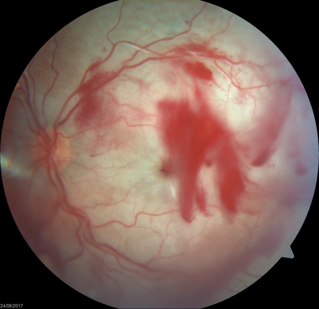
Retinal detachment
Retinal detachments often develop in eyes with retinas weakened by a hole or tear. This allows fluid to seep underneath the retina , weakening the attachment so that the retina becomes detached like wallpaper peeling off a damp wall. When detached, the retina cannot form a clear picture and the
vision becomes blurred and dim. The retinal tissue starts to lose its functionality within 90 minutes of detachment. Hence these symptoms should be brought to the notice of the retina specialist at the earliest so that a timely laser or surgical intervention can restore the vision to the best possible extent.
Retinal arterial blocks
A retinal artery occlusion occurs when the central retinal artery or one of its branches gets blocked. This blockage is typically caused by a tiny embolus (blood clot) in the bloodstream. The occlusion decreases the oxygen supply to the area of the retina nourished by the affected artery, causing permanent vision loss. Patients usually have a sudden loss of vision – typically when they wake up from sleep. Retinal artery obstruction is comparable to a stroke in the eye. The damage can be quite severe, depending on the extent to which the blood flow has been disrupted.People with co-morbidities like diabetes, hypertension , underlying blood disorders are all prone to developing this condition.
Retinal venous blocks
Central and branch retinal venous occlusions occur when there is a congestion to blood flow and an increase in backpressure on the central retinal vein. It causes a variable degree of visual loss and can be easily diagnosed by a retinal examination. It is commonly seen in hypertension and diabetes. Sometimes it can be seen in people with blood clotting disorders. An early OCT scan of the retina can help the retina specialist pick up a macular edema due to the occlusion and can be treated with intraocular injections aimed to salvage the vision to the best possible extent.
Other Disorderstreated are:
- Vitreous hemorrhage
- Cystoid macular edema
- Macular hole
- Retinitis pigmentosa
- Posterior uveitis
- Ocular trauma
- Intraocular tumours
- Central serous retinopathy
- Retinoblastoma
- Hereditary retinal degenerations
- Others
Procedures & Equipment
OPD procedures frequently done
- Indirect Ophthalmoscopy- To have a detailed binocular 3-dimensional view of the retina. The patient is asked to lie down and with an indirect ophthalmoscope, we look at the full extent of the retina. Sometimes, it may be needed to press on the lids to get a good look at the edge of the retina.
- Stereo biomicroscopy- This is done for a magnified examination of the central retina and optic nerve.
- Digital fundus camera and imaging – To take serial pictures to evaluate any subtle changes that may occur with time. A key for all diseases in which we may require observation and follow up.
- Fundus Fluorescein angiography(FFA) and indocyanine green angiography(ICGA)- A dye is injected into the veins of the hand and serial pictures are taken with the Heidelberg ™ imaging apparatus.. This investigation set helps us obtain information regarding retinal and
choroidal blood circulation which is a gold standard for diagnosis of many retinal and choroidal disease states.
- Optical coherence tomography(OCT)- It is one of the most important diagnostic tool to evaluate the depth and extent of almost nearly every retinal condition. It gives us a detailed evaluation of all layers of the retina and choroid similar to a tissue study under the microscope but in a non invasive manner. It is based on reflectance of light waves of a certain wavelength from the retino-choroidal tissues.
- Optical coherence tomography angiography( OCTA) – This investigation helps in understanding the blood circulation of the retina in a non-invasive manner and can help us avoid the FFA/ICGA in certain specific conditions -thus eliminating the need for intravenous dye injection.
At Prabha we make use of the best in class OCT/OCTA and FFA/ICGA imaging apparatus from Heidelberg ™ .
- Ultrasonography and ultrasound biomicroscopy- Standard of care for vitreo-retinal disease diagnosis in hazy media like cataract and vitreous hemorrhage. It is a useful aid in cases of uveitis. An ultrasound biomicroscopy offers view to the periphery of the retina as well as the anterior segment and angle structures.
- Green laser photocoagulation – This is used for treatment of diabetic retinopathy, peripheral retinal degeneration like lattice degeneration, retinal breaks, etc ● Laser indirect Ophthalmoscopy- This is another type of laser delivery system for treatment of peripheral retina, retinopathy of prematurity (ROP), recent post operative cases etc.
Retinal Surgery at Prabha Eye Clinic
At Prabha where quality comes first our surgeons deliver the best of surgical outcomes with India’s first intraoperative real time OCT system equipped with the most advanced ophthalmic microscope in the world, the Carl Zeiss Lumera 700 with Rescan (click here) puts unprecedented accuracy and control in the hands of our surgeons.
We also have the ConstellationTM Vision System, which is an advanced micro incision vitrectomy surgery 23G, 25G, 27G retinal surgery module. It is one of the most efficient machines for retinal surgery.
The Retina department at Prabha makes use of the most accurate instruments of care in performing advanced retinal surgeries like diabetic tractional detachments, vitreous hemorrhage, trauma, retinal detachment, recurrent detachments, macular hole, epiretinal membranes, secondary IOLs, etc .