


The inability of the eye to accurately focus the rays of light coming from a distance on the retina is called refractive error. The correction of the eye to view the image correctly is refractive surgery which again depends on the condition of the eye.
Specialists:
- Dr. Vinay R Murthy
- Dr. Arvind.K
- Dr. Roopashree Matada
- Dr. Kavya N
Refractive corneal surgery:
Refractive surgery wing at Prabha Eye Clinic offers ultramodern approaches to refractive correction that ranges from Contoura, Femtolasik, lasik, ICL and TransPRK (Streamlight).
More About the Different Available Surgeries
- Myopia, or nearsightedness: clear vision close up but blurry in the distance.
- Hyperopia, or farsightedness: clear vision in the distance but blurry close up.
- Presbyopia: inability to focus close up as a result of aging.
- Astigmatism: focus problems caused by the cornea.
More than one refractive error can occur. For eg. myopia and astigmatism
- Glasses are the simplest and most popular way of correcting refractive errors.
- Contact lens is cosmetically better though the maintenance and cleaning of the lenses may be cumbersome and have to be replaced at regular intervals.
- Refractive surgeries are increasingly becoming popular with the advent of more predictable laser treatments.
A variety of treatments are available that correct refractive errors by changing the shape of the cornea.
- PhotoRefractive Keratectomy – Through a process known as Photorefractive Keratectomy (PRK) or EPILASIK –Epipolis Laser In Situ Keratomileusis , mild to high amounts of nearsightedness is corrected using laser light energy produced by the excimer laser. Across the world, over 2,000,000 nearsighted people now enjoy clearer vision having undergone laser vision correction.In this procedure, the laser is applied directly to the cornea according to carefully calculated measurements, without creating a flap. For nearsightedness the relative curvature is decreased; for farsightedness the relative curvature is increased. At the conclusion of the procedure a transparent “bandage contact lens” is placed over the cornea to promote healing. PRK is presently used to treat nearsightedness, nearsightedness plus astigmatism, farsightedness, and farsightedness plus astigmatism.
- LASIK, or laser-assisted in-situ keratomileusis, combines delicate surgical procedures and laser treatment. First surgically a “flap” of corneal tissue less than a third the thickness of a human hair is created the flap to one side is lifted. Laser energy is applied to reshape the cornea according to carefully calculated measurements. When treating nearsightedness, laser is used to decrease the cornea’s relative curvature. When treating farsightedness, the cornea’s relative curvature is increased and then the corneal flap is repositioned. A naturally-adhering bandage, the flap helps to improve results and speed recovery. LASIK may be used for nearsightedness, nearsightedness plus astigmatism, farsightedness, and farsightedness plus astigmatism.
- Femtosecond LASIK – What is FemtoLASIK?
Femtosecond laser creates the flap in less than 15 seconds and the wavelight EX 500 Excimer laser with its advanced iris tracking system corrects the refractive error.
Understanding the FemtoLASIK procedure:
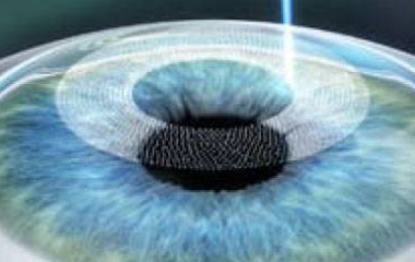
Step 1: A contact lens cone make a gentle contact with the cornea and the FS laser creates a thin flap.
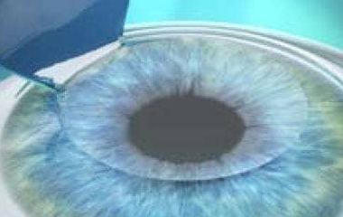
Step 2: The refractive surgeon then exposes the prepared corneal bed for excimer laser treatment by lifting the flap.
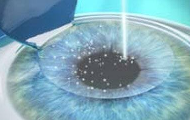
Step 3: The FemtoLASIK procedure is complete when the flap is securely repositioned.
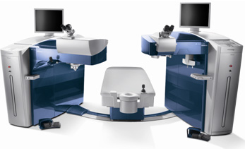
Excimer Laser
Built for Speed and Superb Outcomes
- The WaveLight® EX500 Excimer Laser is the only excimer laser to operate at 500 Hz, with an average treatment time of approximately 1.4 seconds per diopter
- The swivel bed is designed to optimize time between the flap creation and laser procedures Its innovative engineering helps reduce the potential for:
- Stromal dehydration of the cornea
- Flap shrinkage
- Sensitivity to eye movements
- Patient fixation fatigue
Customized LASIK for Every Eye
- Wavefront Optimized treatments
- Customized pupil-center centration options among different lighting conditions
- Line of sight and corneal vertex centration
Precisely Focused on Accuracy
With a powerful 1050 Hz-type multi-dimensional eye tracker, synchronized at 500 Hz, the WaveLight® EX500 Excimer Laser offers safety and exceptional precision:
- Movement tracking with just 2 milliseconds of latency*
- Dynamic pupil tracking from 1.5 mm to 8.0 mm
Optimized Shot Distribution
Utilizing proprietary PerfectPulseTechnology®,theWaveLight® EX500 Excimer Laser maintains a high pulse frequency with a minimized thermal load4,1:
Only 1 pulse in 5 is allowed to overlap
- Optimizes temporal and spatial shot distribution
- Additional pulses are sent to the periphery to compensate for energy loss
- Reduces the potential for nighttime glare
Designed with Efficiency in Mind
The WaveLight® EX500 Excimer Laser workstation has been ergonomically designed to provide enhanced information and usability throughout the refractive procedure:
- Integrated heads-up display
- Fiber slit projector with LED light source
- Increased working distance of 25 cm
- Standard, high-resolution video system
- Built-in, non-contact, online pachymetry
The Cutting Edge of Laser Technology
Featuring enhanced beam path technologies, the WaveLight® EX500 Excimer Laser sets the standard for laser precision and durability:
- Completely sealed beam path
- Continuous nitrogen-flushed beam path
- On board nitrogen generator
- Long laser head life expectancy
With its excellent safety profile, patient comfort and superior visual outcomes, Blade free LASIK is among the fastest-growing refractive surgical techniques in the country today.
The department of Refractive surgeries at our hospital is amongst the very few centers in the world equipped with state of the art and top-line LASIK machines: Femtosecond laser machine, Contoura, and Wavelight EX-500™ which helps us to individualize patient treatment depending upon their suitability. Our specialists at Prabha Eye Clinic will test your eyes completely using the best in class eye equipment and suggest a treatment that suits you the best.
Contoura – “Each Fingerprint is unique, So is your eye”
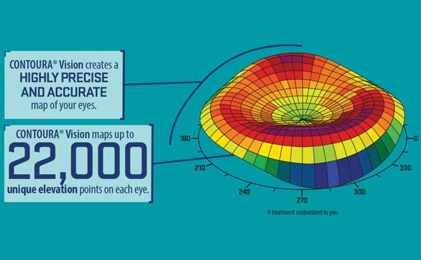
What is Contoura? / Topoguided LASIK ?
Step 1: An initial examination is performed by the refractive surgeon followed by diagnostic tests which includes Pentacam, Tear Film analysis and TopolyserVario.These tests determine the patient’s eligibility and need for Contoura.
The pupil is dilated and retina is checked for areas of thinning. If these are found, a barrage laser is done and Contoura is scheduled after 2 weeks.
Step 2: On the day of the procedure, the scans are reviewed and the refractive surgeon customises the treatment plan and Contoura, FemtoLASIK/ PRK is perfomed with the most advanced refractive platform available in the world, Wavelight Refractive Suite.
Step 3: Femtosecond Laser is used to create a thin flap which is followed by Topoguided customised Laser Ablation using EX500
Step 4: Following the procedure, the patient rests for half an hour. Post operative instructions are explained by a personalised counselling team and the post op checks are done the following day and a month later.
Advantages of Contoura
- Especially suitable for patients with astigmatism
- Gives the sharpest possible vision
- Lesser chances of glare and halos
The department caters to the efficient diagnosis and management of a wide range of diseases:
- Refractive errors management with state of art technology.
- Keratoconus and other ectatic disorders
- Corneal degenerations
- Congential and hereditary corneal disorders
- Corneal infections
- Allergic eye diseases
- Immunologic disorders of cornea
- Dry Eye disease
- Ocular Surface disorders including severe dry eye, blepharitis, chemical injuries, Stevens Johnson’s syndrome, Cicatrizing conjunctivitis and Ocular surface tumors
Surgeries
The department offers most recent advances in the field of corneal surgery which include:
- Collagen Cross Linking for keratoconus using the accelerated cross linking system.
It is the most widely accepted treatment to stop the progression of a clinical entity called keratoconus where, the watch glass of the eye – cornea would have undergone a change in the shape, which may be associated with weakness. During cross linking of cornea, riboflavin– vitamin B2, a photosensitive solution is used to swell up the corneal tissue, following cornea is exposed to UV light of therapeutic range. This procedure is known to strengthen the cornea, however it does not reverse keratoconus.
- Corneal Transplantation
The cornea is the transparent tissue that covers the front of the eye. The cornea is responsible to focus light onto the retina, which is at the back of the eye. Any damage to the cornea results in blurring of vision, watering and other related problems.
A corneal transplant is performed when vision cannot be corrected satisfactorily using other procedures, or when the cornea cannot be repaired or healed with medications. The following conditions may require corneal transplant: corneal decompensation; advanced keratoconus; congenital hereditary endothelial disease; corneal dystrophies; corneal opacity (scarring post infections, post trauma, congenital) non resolving corneal ulcer, impending perforation or perforated corneal ulcer;
Corneal transplantation can done for Optical indications (corneal opacity), therapeutic (non healing corneal ulcers) and tectonic indications (perforated corneal ulcer)
It can be :
1.Penetrating keratoplasty: (full thickness corneal transplantation)
(Optical, tectonic and therapeutic) with anterior segment reconstruction, Combined with SFIOL Vitreo-retinal procedures and Paediatric PK
2. Lamellar corneal surgeries: (Affected part/layer of the corneal tissue is replaced with a healthy corneal tissue)
Including Deep Anterior Lamellar Keratoplasty (DALK) and Descemet’s Stripping Endothelial Keratoplasty (DSEK), Descemet’s Membrane Endothelial Keratoplasty (DMEK)
ICL ie Implantable Collamer Lens
ICL is lens implant that can be used for the correction of refractive errors (myopia, hyperopia, & astigmatism).
ICL is made of bio-compatible material, and it is so soft that it can be injected painlessly into the eye through a tiny opening in the cornea.
Once inside the eye it is positioned in the space between the natural lens & iris. ICL is invisible, ie it is not visible from outside. You can neither see or feel the lens once it is inside the eye.
ICL is supposed to remain permanently inside the eye. However it can be removed easily if necessary, making the procedure a reversible one.
Indications for ICL implantation
- Mild to High Myopia (near sightedness) and / astigmatism –
- Mild to high Hyperopia (far sightedness) and / astigmatism
- Stable Keratoconus (post collagen crosslinking)
- Patients not suitable for LASIK – Thin Cornea, Large pupil, Dry Eye, high myopia and borderline topography
Preoperative assessment
Certain parameters have to be assessed to ascertain eligibility for ICL implantation. These include refraction, corneal topography, corneal thickness, corneal diameter, corneal endothelial cell count ,anterior chamber depth and ultrasound biomicroscopy.
Surgery
Surgery is usually performed under topical anesthesia (eyedrops). The procedure is painless and takes approximately 10 minutes to perform. A few hours later you will be able to leave the clinic and resume most of your activities. Visual improvement is noted immediately post procedure.
FREQUENTLY ASKED QUESTIONS
Am I a candidate for ICL ?
Yes if you have stable nearsightedness( myopia, ie minus power) or farsightedness (hyperopia, ie plus power) and/ astigmatism, adequate anterior chamber depth, normal endothelial count and no other eye disease,
What are the advantages of ICL ?
The lens is very small, foldable, and injected through a tiny, pain free, self healing incision in your eye. ICL provides highly predictable outcomes, excellent quality of vision, and can be removed if necessary.
How quickly can I go back to my daily routine & activities ?
Due to the quick recovery after this treatment, you can leave the center after a couple of hours. You will be able to enjoy your new eyesight almost immediately and go back to active lifestyle . Your surgeon will give you detailed advise.
What if my vision changes ?
Though unlikely if during your annual eye exam major changes in your vision are observed, ICL can be removed or replaced. With ICL you can still wear glasses or contact lenses if necessary. The lens does not treat presbyopia (to check omce as presbyopic ICLs are available now) or eliminate the need for reading glasses due to age.
What is the long term experience with ICL ?
ICL has been available internationally for over 15 years. Millions of lenses have been implanted since then.
Facilities & Diagnostic Services
Corneal Topography (PENTACAM,TMS-4)
Evaluates shape and power of corneal surface. The Pentacam, based on the Schiempflug photography is unique in that it provides invaluable information that other instruments are incapable of measuring.
Pachymetry – Measurement of corneal thickness
Specular Microscopy – Study of the corneal endothelium (cells that maintain corneal clarity)
Anterior segment digital photography
Microbiological and histopathological evaluation of diseases of external eye diseases
Anterion – which is a single workflow-efficient solution that brings together corneal topography and tomography, biometry measurements and IOL calculation to transform the day-to-day routines in cataract and refractive surgery as well as cornea and glaucoma diagnostics. With this station comprehensive measurements are taken while minimizing the time of acquisition.

Topolyser Vario – a topographer which aids in picking up aberrations / irregular corneal surface –which is a promising tool in planning Contoura/ topoguided treatment
Ocular Surface Analyser/ Lipiview – A diagnostic tool to detect the various subtypes of dry eye which enhances decision making in giving targeted therapy to patients affected by dry eye of various etiology