


Glaucoma Services provided at Prabha Eye Clinic include evaluation and treatment of Glaucoma and associated diseases.
Our Glaucoma Specialists are:
- Dr. Gowri J Murthy
- Dr. Priyanka Sudhakar
We manage all types of glaucoma and provide comprehensive clinical examination including Gonioscopy, Tonometry and detailed Fundus evaluation. Investigative modalities that we have include Humphrey Visual Field Analyser, Optical Coherence Tomography, Pachymetry, Ultrasound Biomicroscopy and our newly acquired Anterion for anterior segment imaging.
Laser treatment modalities that we provide include, Nd YAG Laser for peripheral iridotomies, and capsulotomies. We also perform Trans-scleral and Endoscopic Cyclophotocoagulation, Cyclocryotherapy in advanced glaucoma. We offer a wide range of Surgical options from the most conventional to the latest, and include Trabeculectomy, Ahmed Glaucoma Implant and Minimally Invasive Glaucoma Surgeries such as IStent, Kahook Dual Blade(KDB) excisional goniotomy and Bent angle needle goniotomy(BANG). Specialised and complex cataract surgeries in eyes with previous glaucoma surgeries, Pseudoexfoliation, non-dilating pupils, traumatic eyes needing repair and special conditions like nanophthalmos, anterior segment dysgenesis are also done in addition to the standard combined cataract and Glaucoma surgical procedures.
We also extend our services to the pediatric population and offer clinical and surgical management of Glaucoma in newborn, infants and children. Goniotomy and Trabeculotomy are also performed. Continued patient education and availability of patient counselling services helps us in a providing a more holistic approach to patient-care
More Info on Glaucoma
Laser Treatment in Glaucoma – Glaucoma laser surgery is usually done in a clinic and it is painless. During laser, a tiny but powerful beam of light is used to treat a part of the eye .
The following are the most common laser surgeries to treat glaucoma.
LASER PERIPHERAL IRIDOTOMY.
- Used in treatment of narrow angle glaucoma. It makes a small hole in the periphery of the iris and helps to drain the fluid.
- It helps to open the angle and helps in improved drainage of the fluid.
- Reduces chances of extreme fluctuations in eye pressure.
- Prevents sudden increase in eye pressure which can lead to an acute attack of angle closure glaucoma.
How is it done?
- Local anaesthetic drops are instilled in the eye.
- A lens in kept over the eye to focus the laser energy, and to keep the eye still.
- Laser is focused on the iris and a small laser hole is created.
- There will be no pain during the procedure; however, you might feel a small jerky sensation in the eye as the laser is being done.
- The doctor will need you to keep your eyes steady during the laser.
- Post laser eye steroid eye drops will be prescribed.
- Continue all other glaucoma medications being taken by you, unless instructed otherwise.
- Your vision may be slightly blurred on the day of the laser.
CYCLOPHOTOCOAGULATION
This is done to decrease the production of the fluid in the eye by lasering the site of production in the eye, the ciliary body. This procedure is reserved for end stage, and complicated glaucomas. This can be done through the external route, i.e., trans scleral photocoagulation, or by the more precise internal endoscopic route, namely endoscopic cyclophotocoagulation. Endoscopic Cyclophotocoagulation has better profile in terms of vision preservation compared to transcleral Cyclophotocoagulation.
These procedures are not meant for primary treatment of glaucomas ,and are reserved for complicated situations.
It is mainly used to save the eye from severe glaucoma damage not being managed by standard glaucoma surgery
Glaucoma surgery is done to maintain the existing vision in the eye. It cannot improve vision, or restore vision that has been already lost.
What is Glaucoma?
Glaucoma is a group of diseases in which the nerve of the eye, the optic nerve undergoes a slow weakening/ degeneration, leading to loss of visual field. (What is visual field?- visual field is the area seen at one instant by an eye)
Why does one get Glaucoma?
Glaucoma is a disease which is partially genetically determined. If your parents, or siblings have glaucoma, then the chances of your having glaucoma are higher. Apart from these, certain conditions like, steroid use, injury to the eye, certain types of cataracts can predispose a person to develop glaucoma.
How does the doctor suspect that a person may be suffering from Glaucoma?
If during a routine comprehensive eye examination, the ophthalmologist records
- High Intra ocular pressure, high pressure in the eye: Normally the eye pressure is 10-20 mm hg. If the doctor records at least two repeated readings of 22 mm hg or higher, then he/ she is likely to advise a comprehensive evaluation to rule out glaucoma.
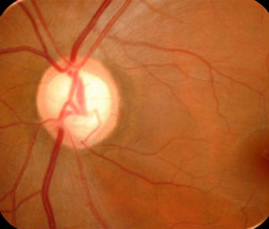
- Difference between the optic nerve head appearance between the right and left eye: Normally, the retina and the optic disc are checked by the doctor doing a routine eye check up. If he/she finds a marked dissimilarity between the eyes, then he advises a comprehensive evaluation to rule out glaucoma. Asymmetry between optic nerve appearances may be a very early sign of glaucoma.
- If the angle of the eye is narrow: If your doctor on routine exam, finds that the junction between the cornea and the iris in your eye, a structure called angle, is narrow, then he may advise a few tests to rule out a type of glaucoma called angle closure glaucoma.
- If you have a very strong family history of glaucoma, and it has caused loss of vision in some of your family members, then the doctor may advise you to undergo certain investigations which serve as baseline recordings, in case you develop/ have a tendency to develop- glaucoma later.
Can I have any symptoms from Glaucoma?
The most common type of glaucoma, called primary open angle glaucoma, at its initial stages, does not cause any symptoms to the patient. There is no pain, redness of the eye, and the patient does not usually notice early damage to the visual fields in one eye.
Glaucoma, there fore is a silent disease, and can be picked up only by a comprehensive eye evaluation.
In angle closure or other acute glaucoma, one can experience pain, in the eyes, redness, seeing colored haloes around lights ,and sometimes headaches. In such situation examination of the angle of the eye by a test called gonioscopy, will alert the doctor to the presence of angle closure glaucoma.
What are the tests which will be done ?
Humphrey visual fields test: This is the current accepted gold standard test to make a diagnosis of glaucoma. In this test you will be required to look at a central light, in a dome. As the instrument randomly flashes peripheral bits of light, you are required to press a button, to indicate whether you have seen the light spot or not. The test takes about 30 mins for both eyes, and may vary depending on the speed of your responses. It is important that you are not tense, or anxious while performing the test, as the results may be affected by anxiety, and inadequate concentration while doing the test. It is perfectly normal to not see any light at least half the number of times, as the instrument is testing at the border of your vision capability.
A technician will administer the test. If you have not understood any aspect of the test, or if you feel uncomfortable during the test, please bring it to the notice of the consultant in charge, or the secretary. The technicians will try and make you comfortable and help you do the test in a calm and unhurried fashion. One of your attenders can also be in the room while the test is done. However, absolute silence must be maintained, and cell phones of the patients and the attender must be switched off during the procedure.
Diurnal variation of Intra ocular pressure:
Our eye pressures have the tendency to fluctuate in a day and between days.Therefore it is important that we obtain a range of pressure readings rather that decide upon the diagnosis based on a single reading. If this particular test called DVs in short form, is advised for you, you will be required to be present at the hospital latest by9:30 AM, and you will be required to be available in the hospital for two hourly pressure recordings till 5:00 PM in the evening. You can bring your own lunch, or use the catering facilitates available.
If you are applying any medications to the eye, these will be continued in the same manner even when the readings are being taken.
If you require facility to rest/ lie down for a while, please inform the secretary, she will make arrangements for a temporary ward/ inpatient facility in the hospital. If you wear contact lenses , weadvise that you should not use them on the day this test is done.
You can read/ watch TV, or engage yourself in any other way, inbetween the readings. At the timings specified in your slip, you can directly go to the doctor’s room and get the eye pressurechecked. There is no need to wait for your name to be called. Thenurse/ patient care worker will assist you in this regard.
Optic nerve/ nerve fibre layer analysis:
Newer methods to accurately map the optic disc contour, and thethickness of the retinal nerve fiber layer are available today, these instruments are very sensitive and can pick up very early glaucoma. However, the results of these tests need to becorrelated with other clinical findings before a diagnosis of glaucoma is made. If the doctor suspects early glaucoma, you maybe advised to undergo these tests. They serve two purposes, one,to detect if any glaucomatous change is present, and two, to accurately document the present position of the optic nerve headso that when you are seen few years from now, if any changeshave occurred they will be evident. Optical coherence tomography OCT would be used to assess the optic nerve for structural changes.
Pachymetry/ central corneal thickness measurement:
Measuring the corneal thickness by a procedure called ” pachymetry”, can help predict the susceptibility to the pressure related damage.
Abnormal test results- what does it mean?
If , by these tests the diagnosis of glaucoma is confirmed, you will probably be prescribed drops, or other medication as will be discussed by your doctor. You will need to regularly get your eye condition monitored every 3 months, as you will be advised.
If test results are normal?
If the results are clearly negative, then you will be reassured and will not require any further tests or follow up specifically for glaucoma. However, please maintain the records of the tests which has been done. Bring these tests to the hospital each time you come for a check up. Also , even though the doctor has currently cleared you of any problem, it may be better to undergoyearly eye examinations, for maintained health of the eye.
If the test results are Borderline?
If the results are borderline, your doctor may elect to observe and see if the results change over time. In this case you will be required to review every 3/ 6 months once as advised by your doctor.
Remember, our eyes are very precious to us, and it is ourresponsibility to take care of them. Problems like glaucoma arevery much treatable, if detected early. Prompt diagnosis andtreatment can help maintain the health of your eye .There is no need to panic if a suspicion of glaucoma has been indicated in your case. Glaucoma can be effectively treated if it is detected, and earlier the detection and treatment, better.
Tips on Instilling Your Eyedrops Properly
Steps For Instilling Eye Drops:
- Start by tilting your head backward while sitting, standing, or lying down. With your index finger placed on the soft spot just below the lower lid, gently pull down to form a pocket.
- Look up. Squeeze one drop into the pocket in your lower lid. Don’t blink, wipe your eye, or touch the tip of the bottle on your eye or face.
- Close your eye. Keep closed for three minutes without blinking.
Optional: Gently press on the inside corner of your closed eyes with your index finger and thumb for two to three minutes (to keep the drops from draining into your throat and getting into your system). Blot around your eyes to remove any excess.